No subscription needed·$12.99 per study·pay only when you submit Pricing →
No subscription needed
$12.99 per study · pay only when you submit

Veterinary X-ray interpretation in minutes, not hours.

Upload your patient's radiographs and medical history, and get a clear, structured radiology report back in minutes. No contract, no monthly fee, no minimums.

Free account in 2 minutesUp to 6 images per studyPay only when you submit
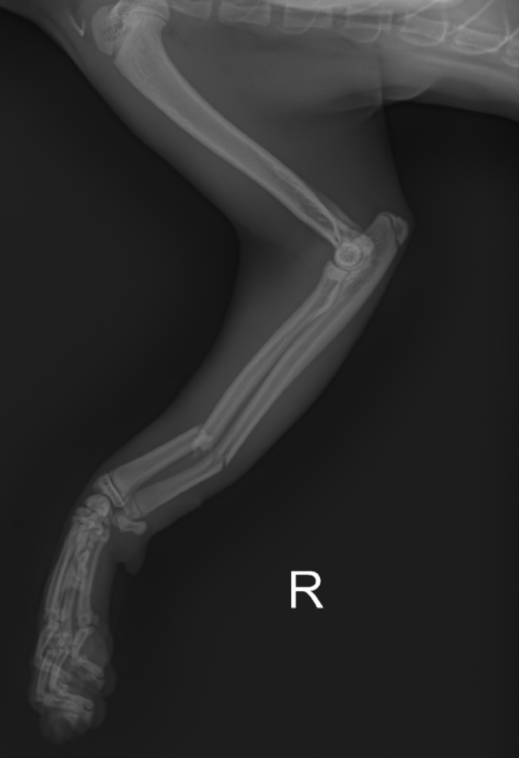
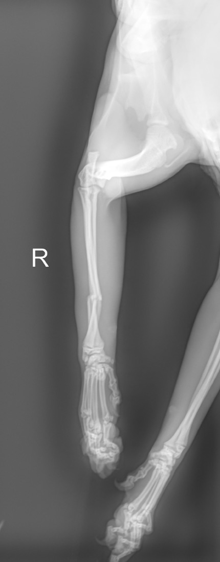
Canine · 1 year · Right forelimbFracture
Lateral radiograph of a dog's right forelimb showing distal radius and ulna fracturesCraniocaudal radiograph of the same forelimb showing displaced, overriding distal radius and ulna fractures
Radius and ulnaComplete distal-third fractures, mildly displaced and overriding
Elbow and carpusCongruent
Growth platesOpen, normal for age
Recommendation: an orthogonal view in the splint to confirm the reduction.

Real case from a partner hospital. Identifying details removed. See the full findings ↓

Three steps. A few minutes.

No software to install, no contract to sign. If you can attach a photo to an email, you can use Rad-Reader.

Create your free account

Your hospital's name, address and veterinarians, so every report is properly addressed. Takes about two minutes.

Upload the radiographs

Up to 6 images per study (DICOM, JPEG or PNG), plus the presenting complaint and history. The history focuses the read.

Pay $12.99, get the report

Your card is charged when you submit. The report arrives in minutes: findings by region, conclusions, recommendations and a PDF.

Get my first read$12.99 per study · no subscription

Real cases, real reports

Two studies from a partner animal hospital, as Rad-Reader reported them. Owner and patient names are removed.

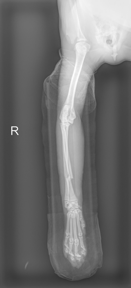
Dog · 1 year · Right forelimb · 3 viewsFracture

Acute, complete, closed-appearing, transverse to short-oblique fractures of the distal third of the right radius and ulna, with mild displacement, angulation and overriding. Residual lateral displacement and overriding of the radial fracture persist in the splint.

  • No radiographic evidence of a pathologic fracture.
  • Elbow and carpal joints congruent; growth plates open, appropriate for age.
  • Probable mild soft-tissue swelling at the fracture site (equivocal).

Recommendation: an orthogonal view in the splint, since reduction can't be fully assessed on a single view.
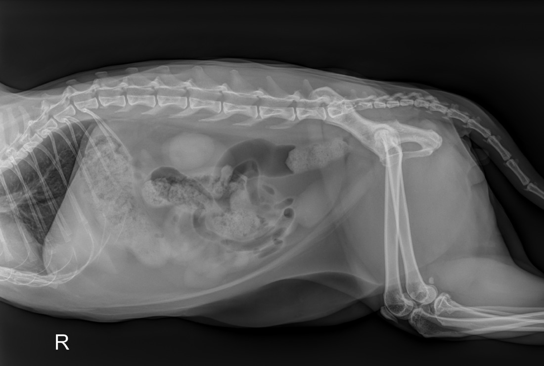
Cat · 8 years · Abdomen · 1 view2 to review

No definite abnormality on this single lateral view. Two items are flagged for the veterinarian rather than guessed: possible prostatic enlargement (the region is poorly defined by bowel superimposition), and the urethra, which is only partly assessable, so calculi can't be excluded.

  • Abundant intra-abdominal and subcutaneous fat: overweight body condition.
  • Granular gastric and colonic contents, likely ingesta and feces; no obstruction.

Recommendation: a ventrodorsal view to complete the urinary tract assessment if there are urinary signs.

Get a read like this · $12.99

Built for busy general practices

A second set of eyes on every film, at any hour, without waiting days or paying for a subscription you don't use.

Minutes, any time

Nights, weekends, holidays. Read while the patient is still in the building.

Several readers, one answer

Independent AI readers review every study; where they disagree, the report flags it for your attention instead of guessing.

A report you can file

Findings by region, conclusions and recommendations, with your hospital and DVM name. Download the PDF for the chart.

Ask a follow-up

Add more history or a specific question ("check the urethra for stones") and the study is read again with it.

View the images anywhere

A full image viewer with window/level, zoom and measurements, from any computer, tablet or phone.

Or connect your X-ray machine

High volume? Send studies straight from your digital X-ray at $9.99 per study, with unlimited image hosting.

Simple pricing. No subscription.

Pay per study, when you submit it. No subscription. If a read can't be completed, you're refunded automatically.

Start today

Pay as you go

$12.99 per study
  • Upload up to 6 images per study
  • DICOM, JPEG or PNG
  • Report in minutes, with PDF
  • Image viewer and follow-up questions
  • Images hosted 30 days (up to 50 studies); reports kept
  • No subscription, no monthly fee
Create free account
For busy clinics with digital X-ray

Connected X-ray machine

$9.99 per study
  • Your X-ray machine sends studies directly (DICOM)
  • Unlimited image hosting while your account is active
  • Just add the history and submit
  • Same report, viewer and follow-ups
  • 50-study monthly minimum ($499.50/month)
How connecting works

Questions

Is there really no subscription?

Yes. Creating an account is free. With pay as you go you pay $12.99 when you submit a study, and nothing else: no contract, no minimums, no monthly fee. (Only the optional connected X-ray plan, for clinics sending studies straight from their machine, has a monthly minimum.)

How fast is the report?

Usually a few minutes after you submit. You'll see the study change from "Reading" to "Read" in your list.

Who reads the radiographs?

Several independent AI readers review every study, following a veterinary radiology checklist, and their results are combined; anything they disagree on is flagged. Rad-Reader is continuously trained by veterinarians: every correction a vet makes to a report is fed back into how future studies are read, so the readings keep improving with use. Rad-Reader is decision support for the attending veterinarian, who remains responsible for the diagnosis. For complex cases we recommend a board-certified radiologist.

What do I need to upload?

Up to 6 radiographs per study as DICOM, JPEG or PNG, the presenting complaint, and any history, exam findings or lab results you have. The more history, the more focused the read.

All questions →

Your next read can be minutes away.

Free account. $12.99 per study. No subscription.

Upload radiographs

Using DVMLog? Your hospital is already connected: sign in with your usual account.